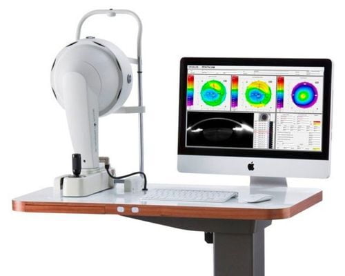
Oculus Pentacam
The Pentacam® measures the entire cornea from limbus to limbus without contact. Use the Corneal Optical Densitometry Display to examine the cornea in detail and to plan your refractive surgery.
Kevin D. Roberts - CEO
Kevin D. Roberts - CEO
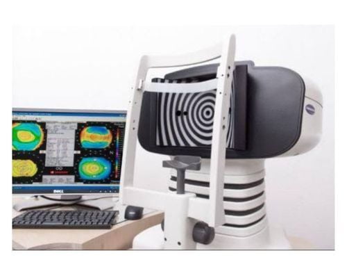
ORB Scan
A topography unit is a machine that takes a specialized photo of the cornea, the front transparent window of the eye. The resulting photo is a topographic map of the corneal surface, similar to satellite aerial colour-coded maps of mountains and valleys on the planet earth.
www.lasikmd.com
www.lasikmd.com
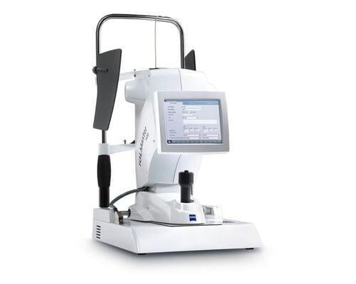
IOL Master Equipment
What is an IOL? An intraocular lens implant is a synthetic, artificial lens placed inside the eye that replaces the focusing power of a natural lens that is surgically removed, usually as part of cataract surgery.
https://aapos.org/glossary/intraocular-lens-implant-iol
https://aapos.org/glossary/intraocular-lens-implant-iol
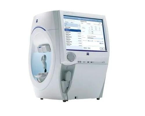
HFA (Humphrey Field Analyser) / Visual Fields
Humphrey field analyser (HFA) is a tool for measuring visual field and is used for detecting monocular visual field but can be used for screening, monitoring and assisting in the diagnosis and monitoring of certain conditions such as glaucoma and brain lesions.
https://terraceeyecentre.com.au/technology/humphrey-field-analyser-hfa/
https://terraceeyecentre.com.au/technology/humphrey-field-analyser-hfa/
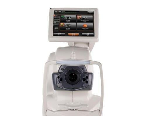
OCT (Optical Coherence Tomography) ONH (Optic Nerve Head)
Optical Coherence Tomography is a noninvasive imaging technology used to obtain high resolution cross-sectional images of the retina. The layers within the retina can be differentiated and retinal thickness can be measured to aid in the early detection and diagnosis of retinal diseases and conditions.
https://ophthalmology.med.ubc.ca/patient-care/ophthalmic-photography/optical-coherence-tomography/
https://ophthalmology.med.ubc.ca/patient-care/ophthalmic-photography/optical-coherence-tomography/
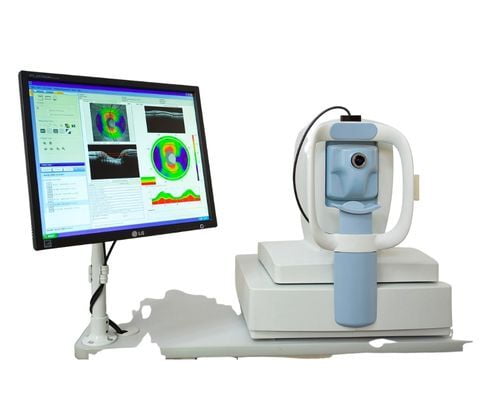
OCT (Optical Coherence Tomography)
OCT-Fundus Camera system that automatically performs alignment, focus and capture with a single touch. The resulting reports enable comprehensive analysis of the macula, optic disc and anterior segment.
https://topconhealthcare.com
https://topconhealthcare.com
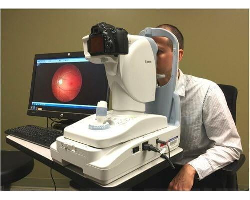
Fundus Imaging
A fundus camera is a complex optical system used for imaging the retina of the eye. Retinal imaging presents a unique difficulty considering that the retina must be illuminated and imaged simultaneously, a process which forces illumination and imaging systems to share a common optical path.
https://www.ncbi.nlm.nih.gov/pmc/articles/PMC2845292/
https://www.ncbi.nlm.nih.gov/pmc/articles/PMC2845292/
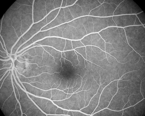
FFA (Fundus Fluorescein Angiogram)
This test is done to see if there is proper blood flow in the blood vessels in the two layers in the back of your eye (the retina and choroid). It can also be used to diagnose problems in the eye or to determine how well certain eye treatments are working.
https://medlineplus.gov/ency/article/003846.htm
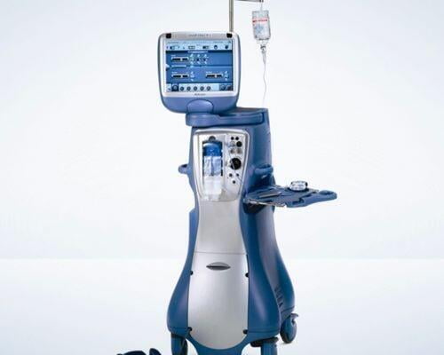
Alcon Legion Phacoemulcification
Phacoemulsification is a modern cataract surgery first developed by Charles Kelman in 1967. The discovery of phacoemulsification came as a boon for the medical fraternity, where a cataractous lens could be emulsified through a small incision of 2-3 mm, giving perfect visual outcomes
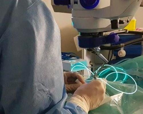
Posterior Vitrectomy
Vitrectomy is a surgical procedure undertaken by a specialist where the vitreous humor gel that fills the eye cavity is removed to provide better access to the retina. This allows for a variety of repairs, including the removal of scar tissue, laser repair of retinal detachments and treatment of macular holes.
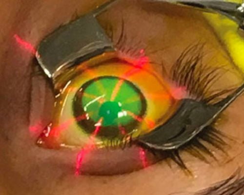
C3R - Corneal Collagen Crosslinking
Corneal cross-linking is a treatment for an eye problem called keratoconus. In this condition, the front part of your eye, called the cornea, thins out and gets weaker over time. This makes it bulge into a cone shape, which can distort your vision and make it hard to see. If the symptoms of keratoconus get severe, you will need a corneal transplant.